Lattice Degeneration
Overview & Our Approach to Lattice Degeneration
Our approach to managing lattice degeneration is comprehensive and proactive, focusing on preventing potential complications and preserving vision. We employ a combination of traditional and integrative therapies tailored to the specific needs of each patient. Our approach includes:
- Strengthening Retinal Health: We use a combination of supplements, acupuncture, and other holistic therapies designed to support overall retinal health and reduce the likelihood of progression to more serious retinal conditions.
- Telehealth & In-Office Treatments: We offer personalized care through telehealth consultations and in-office treatments at our New Jersey office. This allows us to provide timely interventions and closely monitor the condition.
- At-Home Care with ACS3000: For ongoing support, we provide patients with recommendations for at-home care strategies, including lifestyle adjustments and the use of specific devices that promote eye health.
- Targeted Supplements: Our approach includes the use of specialized supplements aimed at maintaining the health of the retina and surrounding tissues, helping to prevent further thinning and degeneration.
Symptoms & Causes of Lattice Degeneration
Symptoms of Lattice Degeneration:
- Typically Asymptomatic: Most patients with lattice degeneration do not experience any noticeable symptoms. The condition is usually discovered during a routine dilated eye examination.
- Peripheral Vision Impact: Although symptoms are rare, some patients might experience issues related to peripheral vision due to the thinning of the retina in areas away from the central macula.
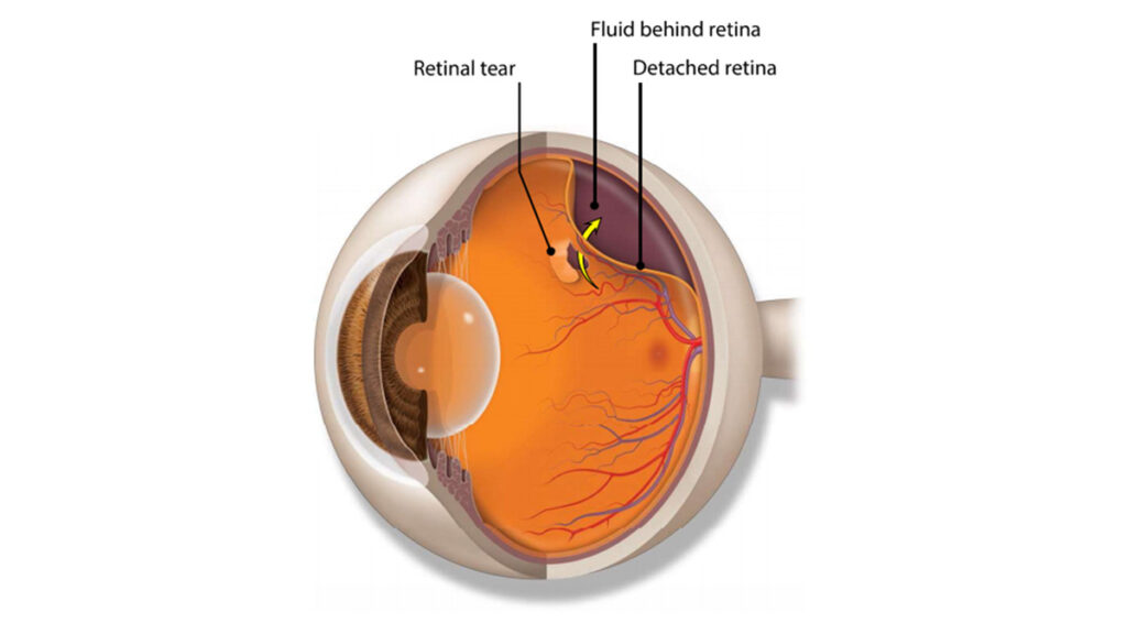
- Increased Risk of Retinal Tears or Detachment: Patients with lattice degeneration have a higher risk of developing retinal tears or detachment, which may present with symptoms such as flashes of light, a sudden increase in floaters, or a shadow or curtain over part of the visual field. These symptoms require immediate medical attention.
Crisscrossing White Lines: The characteristic white lines seen in lattice degeneration are due to retinal blood vessels and fibrous tissue on the retinal surface. These lines are visible upon examination but are present in only about 10% of all lattice lesions.
Lesions and Retinal Holes: Lesions are present in the retina in lattice degeneration, and these can sometimes result in small holes or breaks. Approximately 25–35% of lattice lesions are associated with these atrophic retinal holes.
Uncomplicated Lattice Degeneration: In 90% of cases, lattice degeneration does not involve tears, holes, or breaks. These cases are known as uncomplicated lattice degeneration, where the lesions are stable and do not immediately threaten vision.
Causes of Lattice Degeneration:
- Retinal Thinning and Atrophy: Lattice degeneration occurs when the peripheral retina, which is responsible for peripheral vision, begins to thin, shrink, and atrophy. This can lead to the formation of lesions, small holes, or breaks in the retina.
- Myopia (Nearsightedness): Individuals with myopia are more prone to developing lattice degeneration. The elongated shape of the myopic eye may contribute to the thinning and stretching of the retina, increasing the risk of degeneration.
- Vitreous Changes: As part of the aging process or due to myopia, the vitreous gel within the eye may pull away from the retina, which can contribute to the formation of lattice lesions. However, unlike in retinal detachment, these lesions do not result from traction between the vitreous and the retina.
- Genetic Predisposition: There may be a genetic component to lattice degeneration, as it is more common in individuals with a family history of the condition.

Telehealth and Intensive In-Office Lattice Degeneration Treatment
Start Your Journey with a Telehealth Consultation and Experience the Fastest Results with Our Comprehensive In-Office Care
If you’re seeking the most effective treatment for Lattice Degeneration, our intensive in-office appointments offer the fastest results. Many of our patients choose to visit our New Jersey office for this reason. During these intensive appointments, you’ll undergo a series of advanced treatments tailored to your specific needs over several days, ensuring comprehensive and effective care.
To take the first step toward improved eye health and fast, effective treatment:
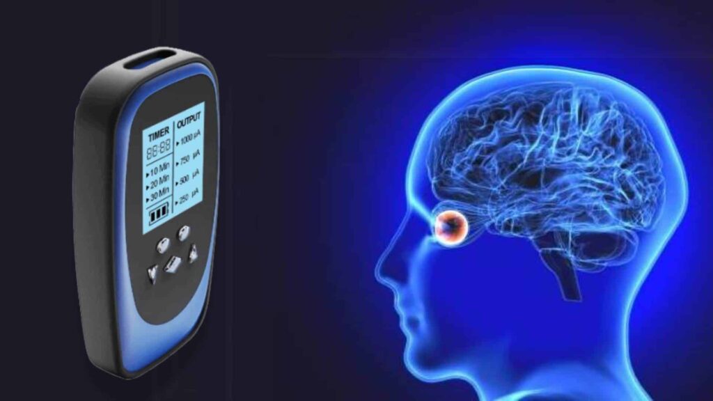
At-Home Lattice Degeneration Treatment with the ACS-3000
Discover the At-Home Frequency Specific Alternating Current Micro Stimulation System
Yes, you can achieve vision recovery from the comfort of your home! Introducing the all-new ACS-3000 at-home vision recovery system. This advanced technology utilizes Frequency Specific Alternating Current Micro Stimulation to support eye health and improve vision.
The ACS-3000 system is designed for convenience and effectiveness, allowing you to benefit from cutting-edge treatments without leaving your home. It’s an excellent option for those seeking ongoing care and maintenance for Lattice Degeneration.
Experience the benefits of the ACS-3000 and see what you’ve been missing. Learn more about how this innovative system can help you!
Shop Lattice Degeneration Supplements
Our team works to create or find the best supportive eye supplements that will give you the greatest effect.
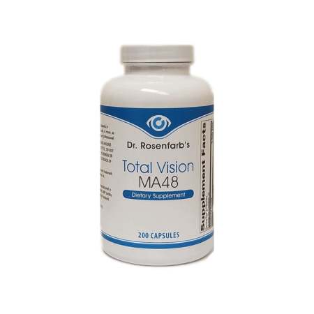
Total Vision MA48
Comprehensive nutritional formula providing the nutrients specifically related to target and support of general eye health and vision.
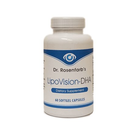
LipoVision DHA

CannaVision
Case Studies
Resources
Join Us on Facebook